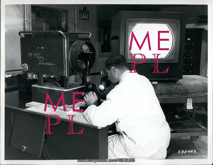
TV microscope for examining patient tissue during surgery
Nov. 11, 1957 - Army Medical Center's TV-Microscope. The US Army announced the development of a colour TV microscope camera now in use at the Walter Reed Army Medical Center which enables the surgeon and the pathologist to examine together tissue removed from a patient during an operation. The tissue is sent from the operating room by pneumatic tube to the Pathology Laboratory, where a slide is prepared and inserted into the microscope. Audio connection permits discussion while surgeon and pathologist examine the specimen. The surgeon can decide whether or not further surgery is necessary before the operation is completed. The assembly allows a three-way connection to include a classroom where students observe the operation, the tissue examination and may ask questions of both surgeon and pathologist. It may also be used solely for laboratory classroom demonstrations and lectures. Developed by Walter Reed and RCA television technicians, the assembly utilises a three-vidicon colour TV camera mounted on a specially designed microscopy bench. Any one of several microscopes can be fitted onto the bench and the camera aligned to view essentially the same field as that seen through the eyepiece of the microscope. Lt. Col Helmuth Sprinz, Laboratory Service, demonstrates a specimen
Pick your License
Restrictions Only for United Kingdom
Aligned Allows Announced Audio Centers Decide Demonstrates Developed Enables Essentially Examination Examine Eyepiece Fitted Further Helmuth Include Inserted Lectures Microscope Microscopes Microscopy Necessary Observe Onto Operating Pathologist Pathology Permits Pneumatic Prepared Questions Removed Solely Specially Surgeon Surgery Technicians Tissue Whether
Features In These Collections
> Contributors > KEY