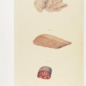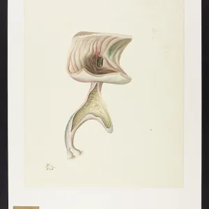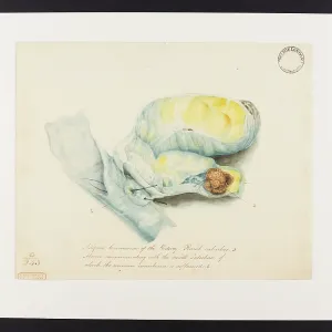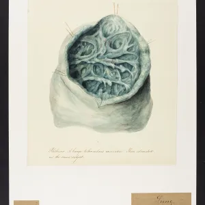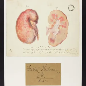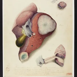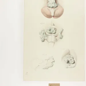
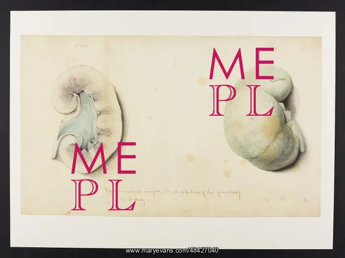
External Surface and Section of Granulated Pale Kidney
Fig I. The external surface. Fig. II. A section of the granulated pale kidney. Hopital de la Pitie, Paris, March 1829
Picture No
13741948
Media ID
48427040
Size
4602px x 3445px
(1.2MB)
Credit
© UCL Library Services Digital Collections / Mary Evans
Source
The Carswell Illustrations Collection - pathological drawings by Sir Robert Carswell (1793-1857), Professor of Pathological Anatomy, UCL Medical School 1831-1840
Date
1829
Pick your License
1829 Anatomical Biomedicine Carswell Granulated Hopital Kidney Pale Pathological Pitie
Features In These Collections
> Contributors > UCL