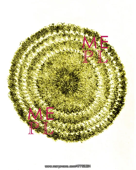
Echinus Spine Section, Early Photomicrograph
Echinus (sea urchin) spine section. Magnification: 60x. Photomicrograph made by Arthur E Smith in the early 1900s, using a combined microscope and camera. In 1904, the Royal Society in London exhibited a series of Smith's photomicrographs to the public. They were later published in 1909 in a book called Nature Through Microscope and Camera. They were the first examples of photomicroscopy many had ever seen
Picture No
13345535
Media ID
47788264
Size
2296px x 2871px
(2.0MB)
Credit
© Science Source / Mary Evans Picture Library
Usage Restrictions
Commercial sales may require 3rd party clearance
Source
Science Source
Pick your License
Restrictions Only for Ireland and United Kingdom
Echinus Micrograph Microscope Spine Spiny Urchin
Features In These Collections
> Contributors > PHR