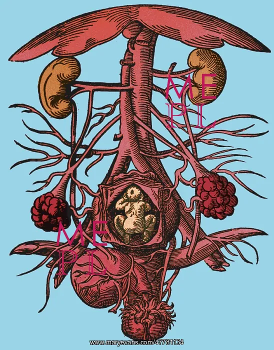
Pregnancy, 16th century artwork
Anatomy of pregnancy. Engraving from the 16th century of the anatomy of a pregnant woman's urogenital system. Within the opened uterus (lower center) is a fetus. The ovaries are at lower left and lower right, with the vagina at bottom center. At bottom left is the bladder, whilst the kidneys are at upper left and upper right. The vertical blood vessels between the kidneys are the aorta artery (narrower of the 2) and the inferior vena cava vein. These split (at center) to form iliac veins and arteries. The umbrella-shaped object (at top) is the liver. Image taken from De conceptu et generatione hominis (1580) by Jakob Rueff
Picture No
13343663
Media ID
47781134
Size
2700px x 3446px
(3.2MB)
Credit
© NLM / Science Source / Mary Evans Picture Library
Usage Restrictions
Commercial sales may require 3rd party clearance
Source
NLM